Sunday, May 11, 2014

Ecology of the Bellarmine Campus


Introduction
In our latest lab, we conducted a survey of the ecosystem--at Bellarmine! Our objective was to track and classify types of organisms based on their ecological function and niche (e.g. primary producer, herbivore, etc.) Through this lab, we learned of the vast diversity of life on campus and how life came together to form an ecosystem. To do this, we simply walked around campus identifying various types of species with our iPads.


Without ado, let's dive into the species we observed around campus!

Species Observed


Producer


Common name: grass
Description: green (indicating of photosynthesis), blade-like
Location found: growing in the quad

Primary Consumer


Common name: chicken
Description: These chickens mainly feed on grain and corn that put them snugly in the place of primary consumers.
Location found: garden


Secondary Consumer
Common name: cat
Description: Sporting retractable claws, this feline hunts for prey such as mice. It rarely attacks adult chickens. 
Location found: dumpster near Bellarmine


Tertiary Consumer 

Common name: human
Description: Able to eat almost anything, humans are at the top of the food chain with no natural predators.
Location found: on campus

Herbivore
Common Name: squirrel
Key traits: Padded paws that allow the squirrel to quickly climb trees and grab fruits and nuts to eat.
Location found: climbing on a tree near the quad

Carnivore


Common name: dog (pitbull)
Description: sharp teeth ideal for tearing meat (not shown), indicating that this species is carnivorous
Location found: sidewalk near Lokey


Omnivore

Common name: crow
Description: with their versatile beaks, crows eat almost anything; from worms, to nuts and seeds.
Location found: Liccardo roof

Decomposer


Common name: earthworm
Description: This little critter that breathes through its skin is actually an earthworm. Its ideal body structure helps it burrow through the soil, feeding on dead organic matter and mixing up the soil!
Location found: ground near the chapel

Pollution Source

Common name: airplane
Key traits: This man made device goes around the campus burning gasoline fuel and serves as a source of noise pollution as well as producing lots of carbon dioxide from the fuel!
Location found: sky above campus

Threatened Species

Common name: grizzly bear
Description: Due to hunting and natural competition, the grizzly bear is going extinct in some parts of the United States, which poses a serious threat to the ecological balance in the Arctic.
Location found: flagpole on the grass

Endangered species


Common name: honey bee
Key traits: This lovable critter that goes around pollinating plants is endangered due to pesticides that cause colony collapse disorder.
Location found: ground near chapel

Non-native species
Common name: Kamchatka horsetail
Description: green (indicating of photosynthesis)
Location found: growing near Liccardo

Discussion questions

1. Define and differentiate between ecology and environmental science and discuss the 
Bellarmine campus in the context of both.

Ecology is a science that seeks to understand the relationship between organisms and their environment, while environmental science is a more general concept that deals with all aspects of the environment--such as biotic, abiotic, chemical, and physical factors. In the context of Bellarmine, ecology would be studying the numerous organisms such as squirrels present on campus and how they affect each other and their environment. However, environmental science would be the study of the complete zone of the Bellarmine campus--such as the soil's nitrogen content, the sources of pollution, etcetera. 

 2. define and describe any population, community, ecosystem, biome and aquatic zone that you 
find on campus; and discuss the biotic and abiotic factors that contribute to that ecosystem.

Population: At Bellarmine, there is a sizable population of a few chickens in the garden. This population thrives because they have ample feed (from humans) and have no external sources of competition. 

Community: In addition to the chickens, a community would be found in the garden at Bellarmine. Many species such as the chicken, the earthworm in the soil, and the roses (photosynthesis) share a habitat and interact with each other constantly. 

Ecosystem: The entire campus of Bellarmine would form a complex ecosystem. Some abiotic factors found in this ecosystem include the rich soil, which has many nutrients and just the right amount of water (which allows for growth of various deciduous trees and a wide variety of foliage), and the strong sunlight year-round, which serves in the benefit for plants carrying out photosynthesis. Some biotic factors affecting the ecosystem would be the predator-prey relationship between numerous species on campus, such as the birds that eat worms; and birds of prey that eat squirrels. In addition, another biotic factor is the issue of human-introduced pollution. 

Biome: From observing the species found on campus, I can conclude that Bellarmine is found in a deciduous forest biome. This comes primarily with the climate on campus (well-defined rain seasons), with the foliage (deciduous trees). In addition, There are more deciduous trees than coniferous trees on campus. 

Aquatic Zone: There is not much water on campus, other than the swimming pool. However, some plants on campus have adapted in order to survive partially being drenched under water, due to heavy rains. 

3. construct and discuss a food chain, a food web, and an ecological pyramid based on the 
trophic levels that you observe.
Food Chain
This picture depicts a food chain. A food chain is simply a linear progression of consumption, beginning from the producer to intermediary consumers. For example, in this food chain, the chicken will eat the grass, but the chicken is in turn eaten by a human. 


This image depicts a food web, a sequence of events that is more complex than a food chain. This food web involves most of the organisms in a given ecosystem. For example, in this food web, the grass is consumed by multiple organisms (squirrel and chicken). In addition, the chicken is then consumed by a cat, but it also can be consumed by a human.
This image is an ecological pyramid. It depicts the relative energy that each organism has at each stage of the food chain. The producers, at the very bottom of the food chain, have the most energy, since they derive it straight from the sun. However, the primary consumers that eat these producers lose some of the energy and have to eat more of these plants because the plants do not have much stored energy. In turn, the secondary consumer and tertiary consumer lose more and more biomass and energy at the top of the pyramid, and need to eat more in order to maintain their metabolism. 


4. investigate and discuss any endangered, threatened, and invasive species on campus.

A large endangered species present at Bellarmine would be the honey bee. The honey bee is in danger of extinction across the nation because of pesticides that cause colony collapse disorder. Yet, bees are extremely important to the balance of the environment--because in places such as on the Bellarmine campus they are pollinators that help spread the pollen of plants to help them reproduce. Another threatened species observed on campus (not exactly physically present) would be a grizzly bear, present on the flag of California that flies near the quad. Because of hunting and natural competition, the grizzly bear is rapidly losing its dominance in the United States. Already, the California grizzly, depicted on the flag, has gone extinct. If the grizzly bear were to be pushed into being endangered, salmon that the bears help keep in check would probably grow more plentiful and throw river ecosystems out of proportion through their rapid breeding and proliferation. 

5. Define pollution, and describe and discuss the various types that you observe on campus.

Pollution is the presence of harmful substances on campus that damage the environment or the organisms present in the environment. One of the largest sources of pollution would be pollution from burning fossil fuels, which generates carbon dioxide--which is toxic to many organisms and contributes to global warming. This occurs on the Bellarmine campus through such man-made objects such as cars and airplanes in the sky. However, there is also sound pollution as well--from things such as the cement plant next to Bellarmine. While it may not be detrimental to most organisms, it is extremely annoying to listen to for humans. 


Monday, May 5, 2014

Fishies!

Introduction
 The lab we conducted was observing how temperature affected the respiration of goldfish. The purpose of this experiment was to study how the body adjusts its functions depending on temperature, and to see how different animals such as a goldfish breathe. Before the lab, we hypothesized that as the temperature increased, the goldfish would take more breaths. In addition, as the temperature decreased, we expected the goldfish to take less breaths. We thought so because heat and chill affects the rate at which body functions occur, such as breathing. To tackle this problem, our methodology was to place the beaker containing the goldfish in water baths of various temperatures, such as adding ice to the bath to make it cold. This way, we could accurately observe the goldfish's breathing rate without killing it. Ultimately, some key findings were that the goldfish's breathing rate did indeed slow down when placed in the cold bath, while it rapidly went up when placed in the warm water bath.


Materials and Procedure

Materials:
(1) goldfish in a beaker with non-chlorinated water
(1) bluetooth temperature probe
(1) bowl
(1) iPad timer
(1) tank of warm water
(1) chest of ice cubes
(4) beakers used to scoop ice cubes/warm water

Procedure

  1. First, we retrieved the goldfish from Mr. Wong's table.
  2. We put the temperature probe in and made sure that it was room temperature.
  3. Then, we used the iPad timer and two group members counting in sync to record the number of breaths at room temperature (23 degrees Celsius) the goldfish took in 5 minutes.
  4. We then scooped several cupfuls of warm water from the warm water tank and poured it into an empty bowl, which we then placed the goldfish in.
  5. After ensuring that the temperature was above 25 degrees, we then counted the number of breaths the goldfish took in this temperature range.
  6. We then changed the temperature of the bath by scooping up ice cubes and dumping them in until the temperature dropped to 14 degrees. 
  7. Again, setting the timer for 5 minutes, we recorded the number of breaths the fish took.
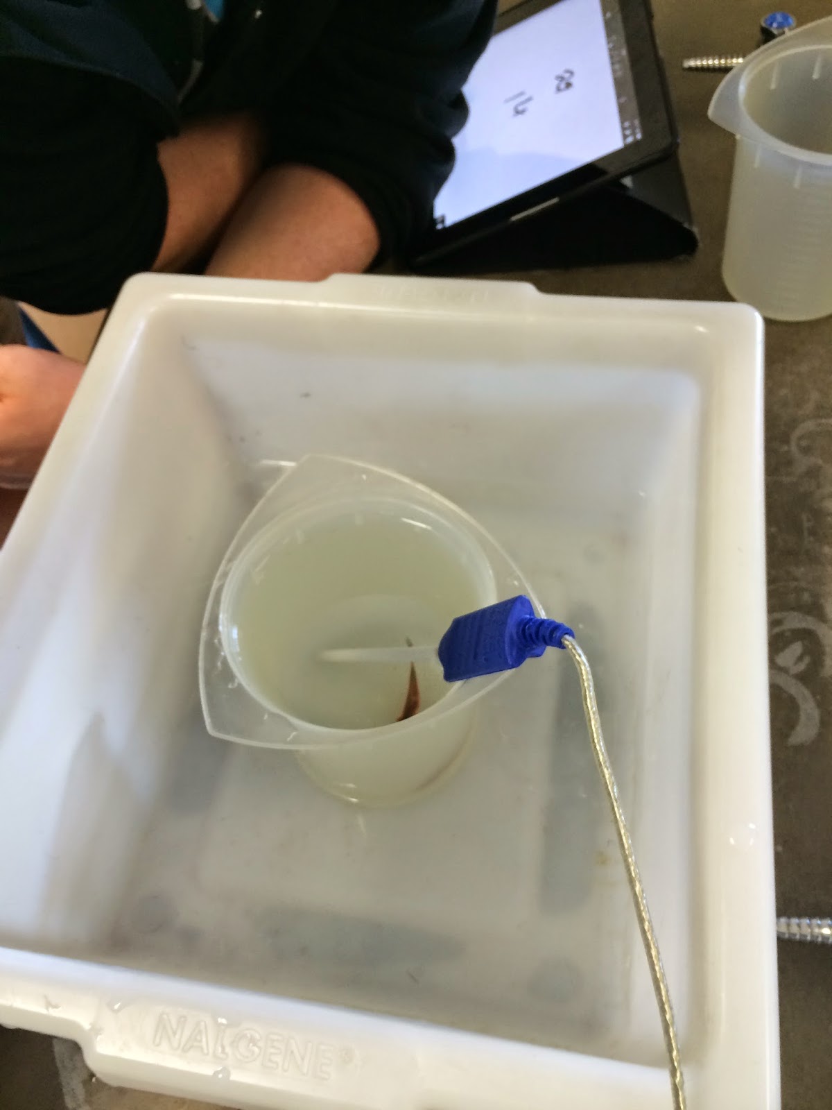
Ice water bath

Data
For this experiment, the room temperature was 23 degrees C (rather than the 15-20 degrees suggested by the manual). In addition, we raised the temperature of the bath to 29 degrees C. Finally, the last temperature used was 12 degrees C. Unfortunately, we did not get to observe the fish at 5-9 degrees C because we ran out of time.

Table 1: Summary of Data


Graph 1: Total breaths taken in 1 minute intervals


Table 2: data relative to other groups

The data shows a relatively linear rate of respiration, as the goldfish's number of breaths taken per minute was relatively the same for each interval of 1 minute. As can be seen from the data, the goldfish had sudden uptick of respiration rate when put in the warm water bath. In fact, the total number of breaths it took in this 5 minute period almost totaled 1000, a large increase from the 644 observed when it was at room temperature. However, when it was placed in the ice bath, the opposite thing occurred: its respiration rate slowed down dramatically. In fact, while the average number of breaths per minute the goldfish took at room temperature was over 100, the average amount of breaths it took in 5 minutes at 12 degrees C was a mere 88.6.

Analysis and Conclusions
From the data, we can safely conclude that the hotter it is, the higher the rate of respiration will be for the goldfish. However, this can also be applied to other animals, such as humans. For example, people who have fever generally have higher respiration rates and heart rates, because of the increase in body temperature. Indeed, our original hypothesis was confirmed by this experiment. One thought as to why this may happen is that as it becomes colder, the goldfish's cellular metabolism dramatically slows down, because of a "numbing" effect that happens, as goldfish are cold-blooded. Therefore, as the metabolism slows to conserve energy, less oxygen is actively needed, and its rate of breathing slows down.  In contrast, as it becomes warmer, the goldfish's metabolic rate increases at a rapid pace. Therefore, its heart needs to pump faster, and in the process, more oxygen is needed in the blood. As a result, the respiratory rate of the goldfish speeds up to help it get this extra oxygen.

1. Describe how the fish's respiration rate is affected by the temperature. Be detailed.
As the temperature increased, the fish's respiration rate increased, from an average of 128.8 breaths per minute at room temperature to 194.6 breaths per minute. In contrast, as the temperature decreased, the fish's average rate of respiration decreased, from 128.8 breaths per minute at room temperature to 88.6 breaths per minute.

2. What other factors (besides temperature) may have affected the breathing rate?
Some other factors may have been that we moved the container (perhaps sending the goldfish into a temporary panic) and the amount of oxygen available in the water.

3. How did your fish compare to the average. Which reading is probably more accurate?
The data we recorded for our fish was slightly higher than the average in each of the three cases (room temperature, cold water, hot water). The reading that is probably more accurate is the average.

4. Why do scientists often take lots of data and look at the AVERAGE? Why do you think you did
 that in this experiment.
Scientists usually take lots of data and look at the average to get the most accurate reading. I think that we did that in this experiment to compare data with other groups and to put a range on our data.

5. Design an experiment that will test how a fish's respiration rate is affected by light. Explain your
 design below.
For this experiment, I would have the control be a fish that is exposed to normal light indoors. I would then change the light levels available to the fish by covering the container with a cloth, to darken it, and move the fish outside or shine a bright lamp on it to increase light levels.

6. Was your prediction at the beginning of the lab correct or incorrect?
It was correct, as the respiration rate did increase as temperature rose.

7. Propose an explanation for your experimental results. Why do you think fish react this way as
 their environmental temperature changes?
Because fish are cold-blooded, their temperature depends on the environment surrounding them. As the environment around them becomes warmer, their metabolism speeds up, and requires more oxygen, which in turn leads to a quicker pace of breathing. 


Monday, April 28, 2014

Physiology of the Circulatory System Lab

Overview
In this lab, our objective was to use a sphygmomanometer to measure blood pressure. We used the sphygmomanometer to listen to the sounds of Korotkoff, and used them to measure systolic and diastolic blood pressure. After measuring the blood pressure, in part 10B, we tested ourselves for our overall fitness. Overall, I expected that with increased exercise, blood pressure would increase (delivering oxygen faster). In addition, I also expected the reclining pulse to be less than a standing pulse because a person would be more relaxed and at ease.

Procedure
Because I did the lab myself, I did not measure the blood pressure of any partner.
1. First, I attached the sphygmomanometer snugly to my upper arm, making sure to secure the wrist strap. In addition, I placed the stethoscope at my elbow.
2. Then, I started pumping air into the sphygmomanometer by squeezing it.
3. After the pressure on the gauge exceeded 200 mm hg, I slowly released air from the cuff.
4. At the same time, I was listening for a pulse. When I heard the first heart sounds, I took down the pressure on the gauge to get my systolic pressure.
5. I continued to release air. When I heard the last sounds, I noted the pressure on the gauge to get my diastolic pressure.
6. I repeated steps 1-5 two more times, and recorded all my data.


Data

Table 1: Blood Pressures
        Trial 1 Trial 2 Trial 3 Average (rounded to closest whole #)
Systolic  111         96         86         98
Diastolic 56         64         60         60

Table 2: Reclining/Standing Blood Pressures
               Reclining     Standing  
Systolic   92                94
Diastolic 57                57
Fitness points = 2

Table 3: A Test of Fitness
Pulse after exercising = 76 (up 8 from 68)
Fitness points = 3

OVERALL FITNESS POINTS = 2+3+3+3+2+3 = 16 (good)

Analysis
Overall, I felt that this lab was extremely fun, as I had the opportunity to learn how to use a sphygmomanometer.  I thought that reading a sphygmomanometer to determine blood pressure was an interesting experience, and enjoyed listening to the sounds of Korotkoff.  In addition, I learned that blood pressure is really a result of the systolic and diastolic cycles of the heart. The heart's contractions cause blood to be pushed into arteries and delivered into various parts of the body, and it is in arteries that you can feel your pulse.  However, I was surprised at my overall fitness. I expected to only be "fair" on the scale for fitness, but ended up earning enough points to score "good."

Answers to Questions

1. Explain why blood pressure and heart rate differ when measured in a reclining position and a standing position.
When measured in a reclining position, a person is much more at ease and feels more relaxed. Therefore, his or her blood pressure is lower than it is compared to standing. When a person stands up, the heart needs to pump harder to support the weight of the person as he is standing up. In addition, standing up requires more energy than normally reclining, which makes the heart pump faster.

2. Explain why high blood pressure is a health concern.
High blood pressure is a health concern because if your blood pressure is too high, it can damage your arteries. For example, a stroke is caused when the blood supply to the brain is interrupted. Therefore, the lack of oxygen means that the brain's function is disrupted, leading to inability to move in most cases. In addition, other parts of the body such as the kidneys, which filter blood, could be damaged if blood does not reach it, which is a grave health concern.

3. Explain why an athlete must exercise harder or longer to achieve a maximum heart rate than a person who is not physically fit.
An athlete must exercise harder to achieve a maximum heart rate because his or her heart is more efficient at pumping blood and distributing oxygen than the heart of a person who is not physically fit. This means that because their heart is used to having exertion, it will take longer for it to pump faster.

4. Research and explain why smoking causes a rise in blood pressure.
Smoking causes a rise in blood pressure because of the active ingredient in tobacco, nicotine. Nicotine is a stimulant that causes the nervous system of humans to go into overdrive. With this, more adrenaline is released by the brain. This causes an increased blood pressure.


Monday, April 7, 2014

Phyla Exam

For this exam, I chose to classify the Otachi Kaiju under the new order repitiliata (reptiles + alata, Latin word for winged), which in itself is under class reptilia.

Taxonomy of the Otachi Kaiju:
Kingdom: Animalia
Phylum: Chordata
Class: Reptilia
Order: Reptiliata
Family: Kaiju
Genus: Quatuor (latin word for 4, because Otachi is a Category IV Kaiju)
Species: Otachi


Characteristics of the Otachi Kaiju:

  • four legs (tetrapod)
  • four-digit wings that can be used to fly
  • spine
  • powerful jaw
  • unique structure: structure on the tongue that is bioluminescent which it uses as a sensory organ
  • sac that can be used to spit a corrosive blue substance
  • tail with three prehensile pincers


The first trait I looked at was that the Otachi has a spine. Therefore, I immediately could classify it as a vertebrate under the phylum Chordata. This is reinforced by the fact that it also possesses a jaw . Because the Otachi is four-legged, I immediately thought of tetrapods. Therefore, I sought to classify the Otachi as a reptile, since reptiles are tetrapods who are also chordates. However, the Otachi kaiju also has wings which it uses to fly. These wings are on the end of four digits, with the fifth digit unfolding the wing when it is in use. Now, I had a dilemma to choose from either reptiles or birds as a class to classify it!

Therefore, I went to a tiebreaker trait. I looked at the unique structure of the Otachi, a structure on the tongue that was bioluminescent and which it seemed to use as a sensory organ. In addition, the Otachi has a sac that can spit a blue acid, similar to the poison of snakes. Therefore, I chose to classify otachi under class reptilia. In addition, beacuse they are tetrapods, I did not choose to classify them as birds. 

For further classification, I created the new order reptiliata (combination of reptilia + ata, Latin for winged). I did this because the kaiju would not solely fit under any existing orders under reptilia, such as the testudines or even squamata, which contain snakes. The family is Kaiju, and because it is a Category IV kaiju, its genus is quatuor (Latin for 4). Finally, its species is simply Otachi. 


The new order reptiliata is meant to consist of reptiles who possess poison or acid glands, and another feature is  heavy wings that allow them to fly. In addition, reptiliata possess a unique tail with three pincers that allow for defense. Howevr, the most important structure of reptiliata is an advanced version of the reptilian tongue that is bioluminescent and allows reptiliata to sense their way, as well as to devour things.

Phylogeny
The main difference that reptiliata have from other orders of reptiles are their wings, and their unique tongue. In this section, I will be exploring the possibilities of evolution for reptiliata.

My theory is that otachi evolved from salamanders.

Wings
The wings of Otachi are located actually on their forearms, which allow it to fly. Therefore, the 
forearms of the Otachi are extremely pronounced. One theory that I can come up with to support the evolution of these wings are that a single Otachi was previously a normal salamander, but due to the excess radiation of the sun, once obtained a mutation that made his arms more fleshy than that of other Otachi and obtained a pouch of flesh near his arms. By stretching the flesh around its arms, it was eventually able to survive better than the Otachi who didn't have the flesh around its arms--beacuse the flesh allowed it to have a thick layer of defense against predators. Therefore, by natural selection, the Otachi who had stretchy flesh developed into wings, survived, and reproduced.
Wings:
http://www.google.com/imgres?imgurl=http://dreager1.files.wordpress.com/2013/07/otachi_2.jpg&imgrefurl=http://dreager1.com/2013/07/&h=720&w=1280&tbnid=Y90W5pe2B93kUM:&zoom=1&docid=mq1Ql8_K0iXW1M&ei=GRxDU8D6GqLhygGi4YBo&tbm=isch&ved=0CDsQhBwwAQ

Tongue
Otachi has a bioluminescent structure on its tongue, that serves as a sensory organ. This tongue probably evolved from the vomeronasal structure found in salamanders, which served the same function and structure (accessory sensory organ). This vomeronasal structure allowed the salamander to have an enhanced sense of smell. However, I believe that radiation from the sun had another effect on this structure. A salamander mutated and had a bioluminescent vomeronasal structure. This allowed it to have more fitness because it could now hunt in the dark better by activating the bioluminescence on will. Therefore, by natural selection, bioluminescent vomeronasal structures were passed on in generations. 

References
http://pacificrim.wikia.com/wiki/Otachi


Monday, March 10, 2014

Pglo Lab Fun!!

Overview

For this lab, our group transformed bacteria using a plasmid, with the objective to make the bacteria glow in the dark. The plasmid contained the gene for GFP (green fluorescence protein), for beta-lactamase, and also contained an inducer that was turned on by arabinose, and triggered production of GFP. In order to quantify our results, we then determined the transformation efficiency of the procedure (how effective our transformation actually was relative to the number of bacteria).

In order to gather our results and data, we observed data such as the amount of growth on each plate, the color of the bacteria, and whether the bacteria glowed in the dark or not.

We expected that the only plate that would contain glowing bacteria was the +lb/amp/ara plate, because it contained lb (to allow the bacteria to grow), and the pGLO plasmid (which helped neutralize ampicillin), and arabinose, which served as an inducer to turn on the GFP gene.

pGlo gene


Materials

E. coli starter plate (1)
Poured agar plates (1 LB, 2 LB/amp, 1 LB/amp/ara) (4)
Transformation solution (1)
LB nutrient broth (1)
Inoculation loops (7) (1 pk of 10)
Pipets (5)
Foam microtube holder/float (1)
Container full of crushed ice (foam cup) (1)
Marking pen (1)

Rehydrated pGLO plasmid (1 vial)
42°C water bath and thermometer (1)
37°C incubator


Procedure

1. First, we used the marking pen to label each tube +pGLO and -pGLO.

2. We then used a new pipet to add 250 microliters of transformation solution to each of the tubes (calcium chloride)

3. After placing the tubes on ice, we used a sterile loop to carefully pick up a single colony of bacteria from our starter plate. We then carefully placed it into the -pGLO tube, spinning the loop until the entire colony was in. We then repeated for the +pGLO tube.
Adding the e. coli

4. We then went over to our instructor Mr. Wong, where he added the plasmid to the +pGLO tube.

5. We then incubated our tubes on ice for 10 minutes. While we were waiting, we examined our plates that were labeled -lb, -lb/amp, +lb/amp/ara, +lb/amp.


6. Using the foam rack as a holder, we put the tubes into a hot water bath (42 degrees Celsius) for exactly 50 seconds. After the heat shock, we immediately transferred the tubes onto ice.

7. After removing the tubes from the ice, we placed them back on the test tube rack at room temperature. We then added 250 microliters of LB nutrient broth to each test tube. Then, we incubated the tubes at room temperature for 10 minutes.

8. We tapped the tubes to mix them. Using new pipettes, we piped 100 microliters from each test tube into their respective plates. We added the contents of the +pGLO tubes to the plates labeled +, and the contents of the -pGLO tubes to the plates labeled -.

9. We then used a new sterile loop to "spread" the bacteria around the plate. We spread them in four quadrants in a zigzag pattern.
Spreading the bacteria

10. We then stacked our plates together and taped them together, and gave them to Mr. Wong, who incubated them for a day.


Data/Explanation of Data


Table 1 


-lb
-lb/amp
+lb/amp
+lb/amp/ara
Color
white
white
white
White
Rate of growth
Lawn of growth
No growth
growth
growth
Glow
no
no
no
yes

As can be seen from Table 1, the only plate where glow was observed was the plate with +lb/amp/ara. The color of the bacteria did not significantly change over the course of transformation. The only place where there was no growth was the -lb/amp plate. We concluded that this occurred because it didn't have the pGLO gene that contained the beta-lactamase gene that would destroy the ampicillin; which was allowed to destroy the bacteria unhindered, and therefore was not resistant to antibiotics. The bacteria in the + plates were antibiotic resistant, and therefore had growth. The LB broth served as a growth medium in this experiment. In the -lb plate, because there was no ampicillin to kill the bacteria, there was a lawn of growth. In the -lb/amp plate, there was no growth even though there was LB because the ampicillin killed it. In the +lb/amp plate, there was growth because LB was present, and the plasmid helped the bacteria neutralize ampicillin, which also occurred in the +lb/amp/ara plate. As we expected, the only plate that contained glow in the dark bacteria was the +lb/amp/ara plate. This occurred because the bacteria was allowed to grow (because of the lb), and the plasmid allowed it to destroy the ampicillin, and the arabinose activated the GFP gene [as an inducer], which produced the protein to make the bacteria glow in the dark. 


Analysis and Takeaways
What I definitely enjoyed most about this experiment was working with the bacteria. I have had almost no experience actually handling bacteria, and this lab allowed me to tackle this experience head on. I particularly enjoyed spreading the e. coli on the plate. My most valuable takeaway from this experiment was a new understanding of transformation and the concept of a gene. We successfully inserted the plasmid, a secondary form of DNA for bacteria. The bacteria glowed in the dark because the pGLO gene produced GFP, which caused the observed change in phenotype. From this lab, I refined my concept of how a gene translate into a protein. In addition, another valuable takeaway from this lab was real life application, because biotechnology nowadays concentrates heavily on the concept of the gene.
 I can begin to see how much easier transformation would be for a large lab. For example, a large lab could possibly easily transform a bacteria to produce insulin, and then bottle and sell the insulin. Reflecting on this experiment, we could have easily increased our transformation efficiency by more carefully completely immersing the plasmid in. In addition, we could have possibly transferred the tubes from the heat bath to ice more quickly. We could have altered this experiment by possibly using other ways to crack open and reseal the plasmid. 

My questions left after this experiment are
Are genetic transformation of animals and human ethical?
How did they calculate the needed times for the heat shock/cold shock? (e.g. 50 seconds and 10 minutes)


Friday, February 21, 2014

Strawberry DNA Extraction

Background
In this lab, our group extracted DNA from strawberries. We accomplished this by breaking open the strawberry nuclei using various techniques and isolating the DNA in a test tube. We then forced the DNA to precipitate.

Materials
strawberries (1)
salt
detergent
gauze (cut into squares)
funnel
ethanol (ice cold)
ziploc bag (1)
pipette (1)
clear stirring rod (1)
paper cup (1)
test tubes (2)

Procedure
1. First, we made the DNA extraction buffer. To do this, we first measured out 45 mL of water in a clear test tube. Then, we measured out 0.75 g of salt by first determining the weight of the paper cup and pouring out salt as needed until the scale read the paper cup's weight plus 0.75 g. We then retrieved 5 mL of detergent in another test tube. Finally, we poured the salt and detergent into the test tube with water to make a 50 mL extraction buffer.
Measuring the salt

Making the buffer


2. After making the buffer, we washed the strawberry and removed its leaves. We then placed it into a ziploc bag, added 10 mL of the extraction buffer, and tightly sealed the bag and all, making sure that no air bubbles were present.


3. We then crushed the strawberries thoroughly for about 1 minute.


Explanation of Steps 1-3
The solution buffer consisted of water, salt, and detergent. The purpose of each was
water: to act as a solvent to the salt, detergent, and extraneous material in the strawberry cells
detergent: to split the fatty acids in the nuclear membranes of the strawberry cells, such that we could breach the nucleus and expose all the lovely DNA. We used detergent because it is nonpolar and could serve this purpose well. 
salt: because salt is an ionic compound (NaCl), it is polar. Because DNA is also polar because of the hydrogen bonding between the nucleotides, salt attracted clumps of DNA together and kept them together.


4. After we crushed the strawberries, we took some gauze and lined the funnel with it, and placed the funnel in another test tube. 

5. Then, we poured the contents of the ziploc bag (the crushed strawberries) through the funnel lined with gauze in order to filter the DNA out from extraneous material. 

6. After discarding the pulp and the gauze, we added an amount of ice cold ethanol equal to the amount of solution left. After swirling it with the stirring rod, the DNA then precipitated and we could observe it on the top of the test tube. (pics below)


The Results!!!!

Cool! Strawberry DNA!
7. Finally, after observing the DNA for a while, we collected it on the rod and observed it.
Strawberry DNA on the rod
Explanation of Steps 4-7
The gauze was employed in order to separate the strawberry DNA from the extraneous material in the strawberry cells. By filtering the contents of the ziploc bag through the funnel lined with gauze, we ensured that all the pulp would not interfere with the DNA that went into the test tube. Furthermore, we used the ethanol to precipitate the DNA. This is because ethanol is more polar than DNA. Because of the difference in solubility between the ethanol and the strawberry solution, the DNA was forced to precipitate. We specifically used cold ethanol so that the enzymes that break down DNA would not work as fast.

Alternative Method(s)
Virtually any alternative method that involves breaking the nuclei of the strawberry open and extracting the DNA using a precipitation reaction can be used. For example, instead of crushing the strawberries, blending might be a better option because it breaks the strawberry down more efficiently, perhaps too efficiently. Nonpolar molecules can be used to breach the fatty acids in the nuclear membranes and open the nucleus. In addition, any polar molecule such as different ions can be used to clump the DNA together, because of the hydrogen bonding. 

Conclusion and Discussion of Genes
From this chapter, we have learned that a gene in reality is just a sequence of instructions for making certain polypeptides. In this context, DNA is really just a molecule that contains the instructions for synthesizing polypeptides. In context of this activity, the DNA that we extracted contains all these instructions to synthesize the proteins necessary for the strawberry. The genes in the DNA that we extracted contains information for synthesizing proteins that carry out functions in the strawberry such as giving it its red tint, forming seeds, and ripening the strawberry.